01
Resolution gap
Conventional CT and MRI can leave clinically important middle- and inner-ear structures below the threshold for confident visualization.
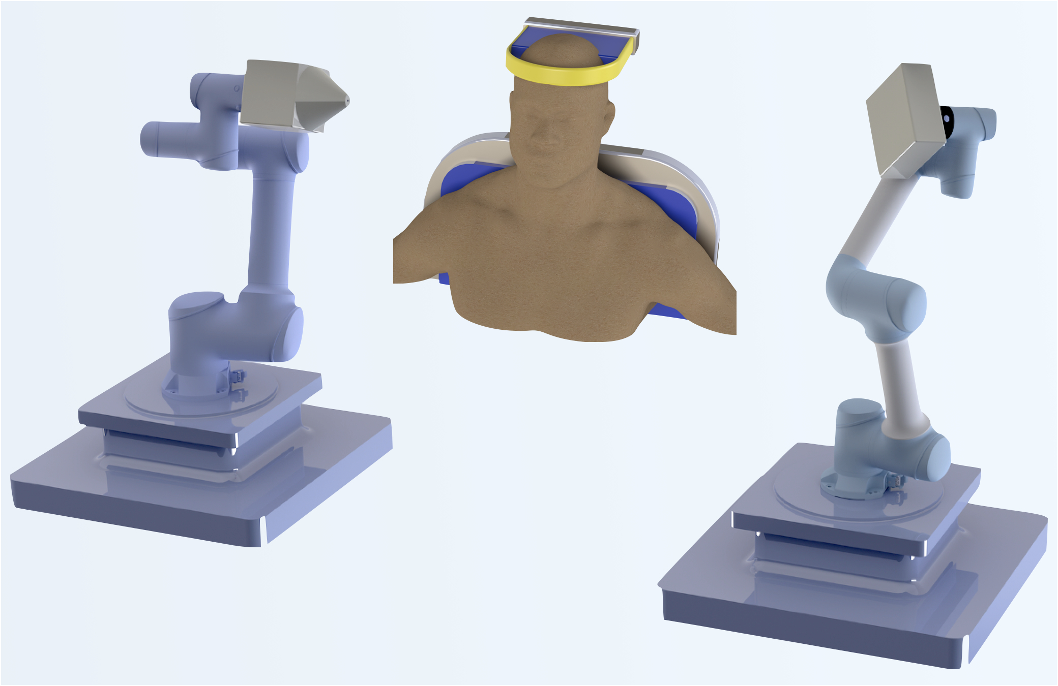
TROI-Imaging is developing a high-resolution region-of-interest CT platform for micro-anatomy—starting with the temporal bone and inner ear, where diagnostic details can approach the scale of hundreds of microns.
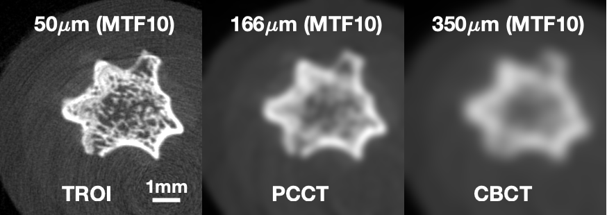
In this comparison, the TROI image resolves substantially finer structures than images simulated to match PCCT and CBCT resolution.
The image shows development-stage technical validation, not clinical performance.
Conventional CT and MRI can leave clinically important middle- and inner-ear structures below the threshold for confident visualization.
Incomplete pathophysiology information can delay diagnosis, limit treatment planning, or push clinicians toward more invasive exploration in operating rooms (ORs).
Concurrent diagnosis and treatment can make OR time lengthy and less predictable, increasing total cost of care. Better pre-operative imaging can help close this gap.
Rather than extending ultra-high resolution across a large field of view, TROI concentrates imaging performance on a clinically relevant region of interest, supporting a compact system architecture and a path toward specialty workflows.
Specialty imaging can create a direct revenue stream within provider workflows, subject to coding, coverage, and payer requirements.
More definitive pre-operative imaging could reduce selected exploratory procedures where anatomy can be resolved non-invasively, making interventions targeted and purely therapeutic.
Better structural information before intervention may improve surgical planning, specialist efficiency, and use of operating-room capacity.
Geometry and acquisition are optimized around compact, high-value anatomy rather than general-purpose whole-body imaging.
Detector performance, focal spot, geometry, reconstruction, motion, and dose must work together to preserve micro-anatomic information.
Cobot-driven design enables free-form trajectories to minimize radiation dose and support flexible patient positioning.
System design, trajectory planning, geometry calibration, and decades of CT reconstruction research form a layered technical moat across the platform.
A platform strategy that could extend into other specialty applications where small-region resolution drives clinical and economic value.
TROI-Imaging brings together complementary expertise in photon-counting CT, image reconstruction, imaging physics, robotic control, and clinical otology.
PCCT expert and inventor on 10+ patents, with deep expertise in engineering and instrumentation. Leads system development and integration.
Clark & Crossan Endowed Chair Professor at RPI and Editor-in-Chief of IEEE TMI; a leading scholar in CT reconstruction and AI-based tomography. Leads reconstruction and AI.
Brings 20+ years of experience in reconstruction algorithms and imaging-physics modeling. Leads physics modeling and imaging simulation development.
Russell Sage Professor and expert in robotics, control systems, and precision motion. Advises robotic control, trajectory planning, and system integration.
Virginia Merrill Bloedel Professor and Director of the Bloedel Hearing Research Center; neurotologic surgeon. Defines clinical requirements and leads clinical validation in patients.
TROI-Imaging is advancing from benchtop technical validation toward a robotic clinical platform. Product configuration, regulatory status, performance specifications, and commercial claims remain subject to development and validation.
We are engaging clinical design partners, strategic collaborators, and investors interested in high-resolution imaging for critical anatomy that conventional systems struggle to resolve.
Contact TROI-Imaging